Back Muscle Chart / Muscles Back Posterior Human Anatomy Vintage Medical Chart. Related posts of back muscles chart muscle anatomy practice exam. They extend and rotate the head and neck. The intermediate layer contains the erector spinae muscles, whose many functions include the extension and lateral flexion of the spine, head and neck. Stylized muscle anatomy chart, front and back. Superficial back muscles, intermediate back muscles and intrinsic back muscles.the intrinsic muscles are named as such because their embryological development begins in the back, oppose to the superficial and intermediate back muscles which develop elsewhere and are therefore classed as extrinsic muscles.
Pain log more pain mapping tools The deltoid, teres major, teres minor, infraspinatus, supraspinatus (not shown) and subscapularis muscles (not shown) all extend from the scapula to the humerus and act on the shoulder joint. The back isn't only one of the body's biggest and strongest body parts, it's also the most complicated in terms of being a series of interconnected muscle groups. For more anatomy content please follow us and visit our website: The intermediate layer contains the erector spinae muscles, whose many functions include the extension and lateral flexion of the spine, head and neck.
Leg Back Muscle Chart by BadFish81 on DeviantArt from pre00.deviantart.net The back isn't only one of the body's biggest and strongest body parts, it's also the most complicated in terms of being a series of interconnected muscle groups. Certain back muscles extend to other areas, like the shoulders, upper arms, and thighs. The trapezius and latissimus dorsi muscles connect the upper limb to the vertebral column. For more anatomy content please follow us and visit our website: The muscles of the lower back help stabilize, rotate, flex, and extend the spinal column, which is a bony tower of 24 vertebrae that gives the body structure and houses the spinal cord. Stylized muscle anatomy chart, front and back. Moves humerus (arm) to chest. Leaning back to straight vertical and all points in between.
We think this is the most useful anatomy picture that you need.
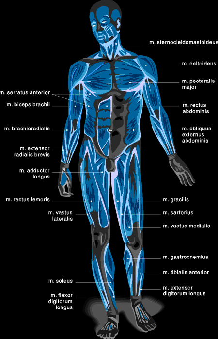
We hope this picture anatomy of back muscles diagram can help you study and research. The superficial group, the deep group, and the intermediate group. This website uses cookies to improve your experience while you navigate through the website. Your clients will thank you for it! Another common cause of lower back and hip pain is disc injury. The rhomboid muscle is activated as you bring and squeeze your scapula or shoulder blades back and together. Muscles found in the superficial group include rhomboid major, rhomboid minor, levator scapulae, trapezius, latissimus dorsi. Superficial back muscles, intermediate back muscles and intrinsic back muscles.the intrinsic muscles are named as such because their embryological development begins in the back, oppose to the superficial and intermediate back muscles which develop elsewhere and are therefore classed as extrinsic muscles. Male body major muscles, flat cartoon vector style infographic illustration. Fit athlete lifting weight with blue muscle light concept on background. Leaning back to straight vertical and all points in between. The deltoid, teres major, teres minor, infraspinatus, supraspinatus (not shown) and subscapularis muscles (not shown) all extend from the scapula to the humerus and act on the shoulder joint. This is a diagram of the larger and more surface muscles of the low back.
However, the spinal erectors travel the length of the entire spine. Both the deltoid and the trapezius are firmly attached to the spine of the scapula. The trapezius and latissimus dorsi muscles connect the upper limb to the vertebral column. The muscles of the back can be arranged into 3 categories based on their location: To download your free copy click the link.
Nerve Chart - Kehoe Chiropractic from kehoechiro.com They extend and rotate the head and neck. Chronic back pain map this tool recommended for: Leaning back to straight vertical and all points in between. To download your free copy click the link. For more anatomy content please follow us and visit our website: This website uses cookies to improve your experience while you navigate through the website. Most of the time, back muscle pain is diagnosed then treated with little more than a prescription of rest, painkillers and muscle relaxants. The superficial group, the deep group, and the intermediate group.
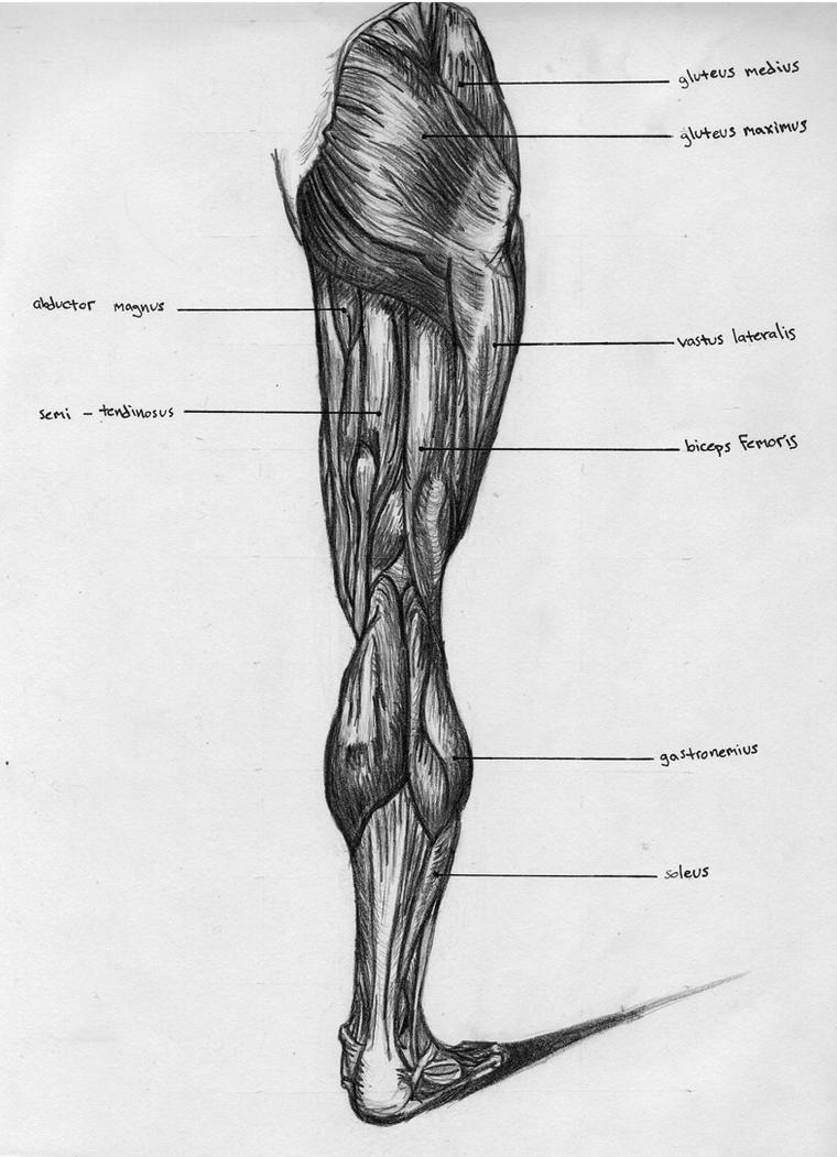
Back muscle diagram back muscles big back big back muscles big lats bodybuilding secrets major back muscles.
The deltoid, teres major, teres minor, infraspinatus, supraspinatus (not shown) and subscapularis muscles (not shown) all extend from the scapula to the humerus and act on the shoulder joint. Claim your free copy of the client back care guide today. We think this is the most useful anatomy picture that you need. Muscles are usually work in pairs because although they can contract and shorten (flex), they are pulled by an opposite (antagonist) muscle to straighten out (extend) again. The muscles of the back can be arranged into 3 categories based on their location: This increases blood flow to the muscle normalizing it and bringing it back to a healthy state. Symptoms of muscle pain include: The trapezius and latissimus dorsi muscles connect the upper limb to the vertebral column. The muscles of the lower back help stabilize, rotate, flex, and extend the spinal column, which is a bony tower of 24 vertebrae that gives the body structure and houses the spinal cord. Another common cause of lower back and hip pain is disc injury. For more anatomy content please follow us and visit our website: Lower back muscle diagram anatomy does degenerative disc disease affect the lower back muscle? Others, like sumo deadlifts, have been shown in emg studies—and in the trenches—to focus more on other muscle groups than the back.
Muscles of the back anatomy. Claim your free copy of the client back care guide today. This is a diagram of the larger and more surface muscles of the low back. Stylized muscle anatomy chart, front and back. People with back pain people who experience headaches printing for use during doctor visits to communicate information about your symptoms quickly tracking your progress over time related tools:
Muscle Chart: Anatomical Muscle Chart - SteroidsLive from www.steroidslive.com The back contains the spinal cord and spinal column, as well as three different muscle groups. Lower back muscle diagram anatomy does degenerative disc disease affect the lower back muscle? Anatomynote.com found anatomy of back muscles diagram from plenty of anatomical pictures on the internet. Another common cause of lower back and hip pain is disc injury. The superficial group, the deep group, and the intermediate group. Muscles of the shoulder girdle posterior. Muscle anatomy ribs 12 photos of the muscle anatomy ribs human anatomy muscles rib cage, muscle anatomy. Others, like sumo deadlifts, have been shown in emg studies—and in the trenches—to focus more on other muscle groups than the back.
To download your free copy click the link.
Both the deltoid and the trapezius are firmly attached to the spine of the scapula. The most common type of back pain is muscle pain—also called muscle strain or soft tissue strain. Some of these muscles are quite large and cover broad areas. To download your free copy click the link. Other muscles are small and cover much less space. Facebook twitter google+ linkedin stumbleupon tumblr pinterest reddit vkontakte share via email print. The rhomboid muscle is activated as you bring and squeeze your scapula or shoulder blades back and together. Related posts of back muscle chart muscle anatomy ribs. Leaning back to straight vertical and all points in between. There are three different muscle groups found in the back: Moves humerus (arm) to chest. The superficial group, the deep group, and the intermediate group. Certain back muscles extend to other areas, like the shoulders, upper arms, and thighs.